Atlanta Breast Wellness 
Thermography Atlanta
Advantage BodyCare Solutions
(770) 355-8352
Call to Schedule an appointment

Introducing the First
Pain-free, Radiation-free
Digital Breast Exam


Digital Infared Thermal Imaging

SureTouch Breast Exam

Lymphatic Therapy


Check, HSA Card, Debit Card, or Zelle

All cards (3% service fee)

(if you require other forms of payment please let me know)

ANNOUNCEMENT

Good News

I Have moved back to Atlanta.

As of  September 1, 2021

I will be providing services in Atlanta/Roswell. 

         500 Sun Valley Dr.     Suite D-1                                                                                                   Roswell, GA.  30076

 It has been my pleasure serving the Atlanta community for the past 20 yrs, And. I am please to announce my return on a full time basis to provide services in the Atlanta area.

Please reach out to me when you are ready to schedule your appointment.

If you have any questions, please contact me by phone, text or email. 

 BLESSINGS and Thank You for your loyalty.

 Dr. Rosalind Gamba, NMD,   

 Phone:  c- 770-355-8352   email:  roz158@bellsouth.net  

What is Breast Thermography?


Breast thermography is a 15 minute non invasive test of physiology. It is a valuable procedure for alerting your doctor to changes that can indicate early stage breast disease.

The benefit of breast thermography is that it offers the opportunity of earlier detection of breast disease than has been possible through breast self examination, doctor examination or mammography alone. 

Thermography can detect the subtle physiologic changes that accompany breast pathology, whether it is cancer, fibrocystic disease, an infection or a vascular disease. Your doctor can then plan accordingly and lay out a careful program to further diagnose and /or MONITOR you during and after any treatment.

Normal


Good thermal symmetry with no suspicious vascular patterns or significant thermal findings.

Fibrocystic Changes


The very significant vascular activity in the left breast justified clinical correlation and close monitoring which returned an opinion of fibrocystic changes taking place. These changes can be monitored thermographically at regular intervals until a stable baseline is established and is reliable enough for annual comparison.

Early Stage Malignant tumor


This is the specific area of a small DCIS. We can see the vascular feed and the discreet area of hyperhermia that is displacing the surrounding hypothermia.

Thermography is a painless, non invasive, state of the art clinical test without any exposure to radiation and is used as part of an early detection program which gives women of all ages the opportunity to increase their chances of detecting breast disease at an early stage. It is particularly useful for women under 50 where mammography is less effective.

Thermography's role in breast cancer and other breast disorders is to help in early detection and monitoring of abnormal physiology and the establishment of risk factors for the development or existence of cancer. When used with other procedures the best possible evaluation of breast health is made.

This test is designed to improve chances for detecting fast-growing, active tumors in the intervals between mammographic screenings or when mammography is not indicated by screening guidelines for women under 50 years of age.

All patients thermograms (breast images) are kept on record and form a baseline for all future routine evaluations.

This patient's thermograms have remained stable for two years. These patterns are like a thermal fingerprint which will only change if pathology develops.


Baseline

3 month Follow-up


First Annual

Second Annual


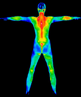
With the new ultra-sensitive, high resolution digital infrared cameras available today a technology that has been developing over the past 20 years is now becoming more accessible.

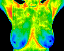
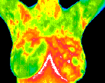
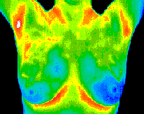
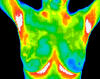
Thermography as a physiologic test, demonstrates heat patterns that are strongly indicative of breast abnormality, the test can detect subtle changes in breast temperature that indicate a variety of breast diseases and abnormalities and once abnormal heat patterns are detected in the breast, follow-up procedures including mammography are necessary to rule out or properly diagnose cancer and a host of other breast diseases such as fibrocystic syndrome, Pagets disease, etc.

Canadian researchers recently found that infrared imaging of breast cancers could detect minute temperature variations related to blood flow and demonstrate abnormal patterns associated with the progression of tumors. These images or thermograms of the breast were positive for 83% of breast cancers compared to 61% for clinical breast examination alone and 84% for mammography.

By performing thermography years before conventional mammography, a selected patient population at risk can be monitored more carefully, and then by accurately utilizing mammography or ultrasound as soon as is possible to detect the actual lesion - (once it has grown large enough and dense enough to be seen on mammographic film), can increase the patient's treatment options and ultimately improve the outcome.

It is in this role that thermography provides its most practical benefit to the general public and to the medical profession. It is certainly an adjunct to the appropriate usage of mammography and not a competitor. In fact, thermography has the ability to identify patients at the highest risk and actually increase the effective usage of mammographic imaging procedures.

Until such time as a cure has been found for this terrible disease, progress must be made in the fields of early detection and risk evaluation coupled with sound clinical decision making.

Thermography, with its non-radiation, non-contact and low-cost basis has been clearly demonstrated to be a valuable and safe early risk marker of breast pathology, and an excellent case management tool for the ongoing monitoring and treatment of breast disease when used under carefully controlled clinical protocols.


Meditherm